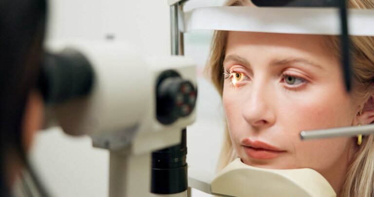
Non-invasive eye tests could improve stroke prediction models by analyzing retinal density, complexity and curvature.
Study: Retinal vascular imprints predict incident stroke: findings from the UK Biobank Cohort Study. Image credit: PeopleImages.com / Yuri a / Shutterstock.com
In a recent study published in Heartresearchers are identifying how specific characteristics of retinal vessels may help predict a person’s stroke risk.
Importance of this topic
Each year, approximately 100 million people experience a stroke, 6.7 million of whom will die as a result of this event. About 90% of strokes are due to hypertension, dyslipidemia, smoking, and unhealthy diet, all of which are modifiable risk factors that respond to lifestyle changes and medication.
The vasculature of the brain has many features in common with cerebral vessels. In fact, a funduscopic examination of the retina thus reveals the health of the vascular bed of the brain.
Previously, the prognostic potential of retinal vascular changes has been investigated. Within the retinal vasculature, tortuosity, arteriovenous ligature perforation, microaneurysms, and venous diameter may reflect the adverse effects of diabetes, high cholesterol, and hypertension.
Despite these observations, mixed results have been reported. Furthermore, current blood tests for predicting stroke risk are expensive, invasive and not highly accurate, thus highlighting the need for better models for predicting stroke risk.
About the study
Data for the current study were obtained from the UK Biobank fundus imaging database with retinal vessel measurements obtained from the Retinal-Based Microvascular Health Assessment System (RMHAS). Data from a total of 45,161 people followed for 12.5 years were included in the analysis.
RMHAS uses an algorithm to analyze 118 measurements while separately evaluating arteries and veins inside and outside the macular area. After adjustment for known risk factors, data were analyzed to identify associations with new stroke.
Conventional risk profile
There were 749 new strokes in the cohort during the study period. Stroke was more common among older adults, men, and participants who were current smokers, as well as those with a history of diabetes. Average body mass index (BMI), blood pressure (BP), and high-density lipoprotein (HDL) cholesterol levels were higher among stroke survivors.
New retinal factors associated with stroke risk
A total of 29 new retinal factors were associated with the risk of new stroke. Seventeen of these parameters were related to vascular density, while three, eight, and one were related to diameter, complexity, and tortuosity, respectively.
Increased risk of stroke with reduced density
For each of the density parameters, one standard deviation (SD) change was associated with an approximately 10–20% increased risk of stroke. Reduced vascular density increased the risk of stroke, which may be due to hypoxia and reduced nutrient supply.
Reduced caliber and complexity increase risk
With caliber parameters, each SD change increased the risk by 10–14%. Reduced diameter relative to length increased the risk of stroke in affected patients.
Parameters related to vascular complexity included fractal dimension, number of bifurcation and bifurcation points, and number of terminal and non-terminal points in arteries. A one SD decrease in complexity was associated with a 10-20% increased risk of stroke.
Of the 30 tortuosity indices, a decreased arterial bending number by one SD increased the risk of stroke. Reduced complexity and tortuosity suggest poor endothelial function, reduced perfusion, inadequate collateral circulation, and increased risk of hypoxia. In these cases, the brain is more easily damaged by stroke risk factors and microbleeds are more common.
Risk models
Compared to using only conventional risk factors, incorporating these retinal vascular parameters resulted in a significant increase in the model’s predictive ability to 75% compared to 74%. When combined with age and gender alone, the vascular model returned performance comparable to traditional assessments, while avoiding the need for invasive blood tests.
Since this non-invasive technique can be combined with age and sex, which are easily obtainable data, this model is a practical method that can be used to assess eye disease and new stroke risk in poor settings. resources and from primary care physicians.
conclusions
Retinal vascular analysis, a noninvasive screening approach to stroke risk assessment, outperforms traditional risk stratification models.”
The current study highlights the importance of density parameters as sensitive markers of stroke risk and offers a novel application of this technology in high-risk individuals. However, additional studies are needed to investigate the role of retinal vascular 29 factors in the pathophysiology of stroke to improve preventive strategies and potential treatments.
Journal Reference:
- Yusufu, M., Friedman, DS, Kang, M., et al. (2024). Retinal vascular imprints predict incident stroke: findings from the UK Biobank cohort study. Heart. doi:10.1136/heartjnl-2024-324705.
