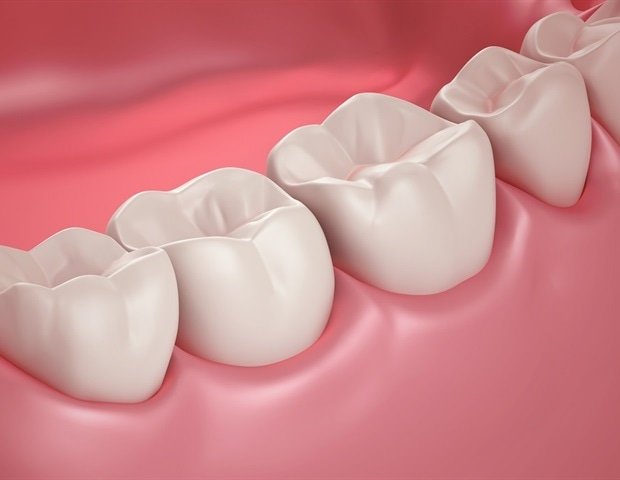
A new study on the natural coordination of tooth development in time and space, led by Dr. Han-Sung Jung at Yonsei University College of Dentistry, Korea, discovered that “lingual” cells on the side of the tongue form the tooth, while those toward the cheek, called “buccal cells,” form the bones and gums, guided by a WMP molecule. These ideas could shape future methods for regenerating, replacing and repairing teeth.
Tooth development is a dynamic process that includes the bud, cap and bell stages, followed by root development and subsequent tooth formation. Processes such as the bud-to-cap transition are mediated by epithelial-mesenchymal interactions. Furthermore, the location of a cell in a developing embryo determines its fate due to relative differences in the concentration of signaling molecules and growth factors.
Scientists have long known that a single tooth develops as a small bud of outer “epithelial” cells within deeper “mesenchymal” cells. It then curves to form a cap shape and then folds further to form the bell shape of a mature tooth, with surrounding bone and gum. Dr. Han-Sung Jung and his team at Yonsei University College of Dentistry, Korea, expanded on these findings by examining how the location of young dental epithelial and mesenchymal cells would affect their development, and published their findings in International Journal of Oral Science.
Lead author Dr. Jung said his team “performed this study to determine how positional identity along the glossobuccal axis determines the distinct developmental fates of the dental mesenchyme. This research has the potential to significantly impact our understanding of tooth development“, he says.
The researchers separated mesenchymal cells on the lingual and buccal sides at both cap and bell stages of a developing mouse embryo and compared gene expression profiles via RNA-seq followed by gene ontology enrichment analysis to understand differences with position and time. They then transplanted the cap-stage tongue and mouth cells separately under the kidney capsule of immunocompromised mice to see what each grew into. The analysis showed that the cells on the lingual side were mainly oriented towards building the tooth itself and shaping its structure, while the cells on the buccal side were more focused on stem cell activity, forming surrounding tissues and supporting tooth growth and repair. Not surprisingly, only the tongue cells in the mouse kidney developed into tooth enamel.
The researchers also reported random mixing of cap stage, oral and tongue cells of genetically modified mice. “We were curious to know if they could find their original location and reorganize when fluorescent lingual and buccal mesenchymal cells were randomly mixed, which they not only did, but the lingual cells grew into dentin to form the tooth as before. This phenomenon is called cellular self-organization,” says first author Eun-Jung Kim.
In addition, they have extensively studied the signaling molecules in each group and found that WNT signaling and R-spondins (Rspo1/2/4) are enriched in lingual cells, along with high proliferation, low cell death and a higher migration rate, aiding tooth formation. On the other hand, oral cells show increased expression of BMP inhibitors, lower proliferation, higher apoptosis and slower migration, favoring the formation of bone and surrounding tissue.
In conclusion, the authors proposed a model of dental cell positioning based on the glossobuccal axis for the formation of teeth and surrounding tissue. Dental mesenchymal cell characteristics were found to vary along this axis, and the fate of tooth and surrounding tissue formation is determined by mesenchymal cells through WNT/BMP signaling. A deeper understanding of the molecular nuances of tooth development will inspire further research in tissue engineering and regenerative medicine, which may ultimately lead to advances in stem cell-based tooth regeneration and more effective therapeutic applications for dental restoration and repair.
Source:
Journal Reference:
Kim, E.-J., et al. (2025). Predetermined dental mesenchymal cells to build a tooth. International Journal of Oral Science. doi.org/10.1038/s41368-025-00391-7
