In a recent study published in the journal Nature, scientists in the United States report the spread of highly pathogenic avian influenza (HPAI) H5N1 virus in cattle in various regions of the United States (US). They further document the detailed symptomatic effects of the resulting disease in these cattle populations. Finally, they use a multidisciplinary approach incorporating epidemiological and genomic analyzes to highlight that virus evolution provides the potential not only to allow cow-to-cow transmission but also efficient multidirectional spread between species, infecting birds, domestic cats, and even and a raccoon. proximity to sick cattle.
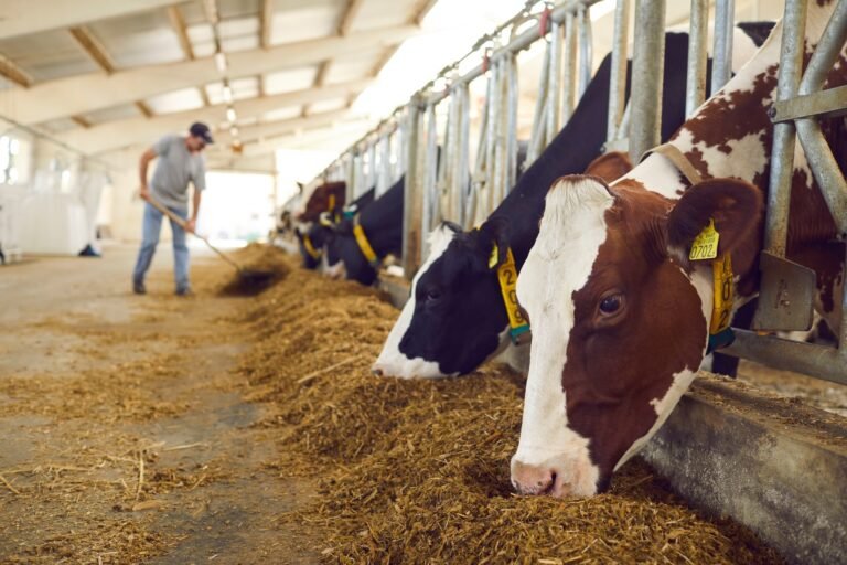
Study: Dissemination of H5N1 highly pathogenic avian influenza virus in dairy cattle. Image credit: Studio Romantic / Shutterstock
Record
Influenza A virus (IAV) H5Nx is a highly pathogenic avian influenza (HPAI) virus that causes widespread respiratory disease and subsequent death in avian populations throughout Africa, Asia, Europe and more recently North America. First discovered in China in 1996, the colloquial “bird flu” has since evolved into eight clades and three neuraminidase subtypes, with the H5N1 2.3.4.4b subtype being its most widespread and epidemiologically relevant agent.
HPAI H5N1 is of concern given the potential for spillover (cross-species infectivity). It has been reported to be transmitted from infected poultry populations to wild birds (2002), mammals (domesticated and wild), and even humans (2003). The World Health Organization (WHO) has recorded 860 human infections and more than 430 deaths since 2003 (mortality rate ~52.8%).
The virus poses significant ecological, economic, and public health threats, having killed more than 90 million birds in the United States (US) alone. The most recent morbidity event associated with H5N1 was in dairy cattle in Texas (TX), New Mexico (NM), Kansas (KS), and Ohio (OH) between January and March 2024. Understanding Epidemiologic and Genomic substrates of this event may allow researchers to elucidate the etiology (origin) of the disease and prepare for future outbreaks.

Influenza A (H5N1/Bird Flu) Influenza A (H5N1/Bird Flu) virus particles (round and rod-shaped, red and yellow). Creative composition and coloring/effects by NIAID. Transmission electron micrograph images courtesy of CDC. Scale modified/not to scale. Credit: CDC and NIAID
About the study
This study documents the incidence of morbidity from January to March 2024 in US cattle across TX and its neighboring states. It uses a detailed interdisciplinary approach that integrates clinical, epidemiologic, and phylogenetic investigations to elucidate the pathophysiology of the virus and the genetic underpinnings of viral dispersibility.
The researchers initially obtained samples for the clinical-epidemiologic evaluation from nine farms in the affected states – TX (5 farms), NM (2), KS (1), and OH (1). Specifically, the only farm in OH was affected after cattle (assumed to be healthy) were imported from the first affected TX farm.
Data collection included nasal swabs, milk, dialysis pads, and serum (n = 331). These samples were subjected to real-time reverse transcriptase polymerase chain reaction (rRT-PCR) and viral metagenomic sequencing. In addition, tissue from birds (big-tailed, rock pigeons) and mammals (cats and raccoons) found dead on contaminated farms were subjected to rRT-PCR analysis.
Virus shedding studies were conducted to elucidate the source and duration of virus transmission after initial infections. The excised tissues from cows, dead birds and mammals were subjected to histological examinations. Finally, phylogenetic analyzes were performed to isolate the causative source of the viral strain and the genetic underpinnings of its significant spread.
Study findings
Clinical-epidemiological investigations revealed multiple disease symptoms in cattle, mainly reduced feed intake, mild respiratory distress, reduced rumination time, lethargy, dehydration, abnormal faeces and abnormal milk production (20-100% reduction in quantity, yellow color and thick consistency ). Symptoms persisted for 5-14 days. However, milk production remained reduced for up to four weeks.
All investigated rRT-PCR samples detected positive viral load, but viral shedding was the highest and most frequently detected in milk and mammary gland tissue samples. Specifically, while virus shedding studies detected viral loads in milk samples on days 3, 16, and 31 post-infection, shedding of infectious virus was observed only on day 3.
“Histological examination of tissues from affected dairy cows revealed marked changes consisting of neutrophilic and lymphoplasmacytic mastitis with apparent obliteration of the tubular gland architecture that was filled with neutrophils admixed with cellular debris in multiple lobules in the mammary gland. The most pronounced changes in his cat The tissues consisted of mild to moderate multifocal lymphohistiocytic meningoencephalitis with multifocal areas of parenchymal and neuronal necrosis.’
Phylogenomic analysis revealed that all recovered viral sequences align with a new monophyletic H5N1 reassortant subtype called B3.13, which was first discovered in a Canada goose in Wyoming (January 25, 2024). This lineage was most closely related to a sequence obtained from a dead skunk in NM (23 Feb 2024). The similarity between the viral genomes from the examined farms highlights circulation and cross-contamination between their inhabitants, possibly due to the transfer and introduction of animals between these farms.
conclusions
The present study highlights the potential for H5N1 virus spillover and cross-infection in avian and mammalian hosts on US farms. The mammary gland was identified as the site of highest viral replication, with contaminated milk representing the most likely route of transmission. The new substrate (B3.13) identified here is of concern given the potential for spread (in domestic and wild bird populations, and even other mammals – cats and raccoons).
Although no human infections were reported from non-study farms, mild infections were reported during the study from other farms near the study area, highlighting the zoonotic potential of the virus and the possibility of a human pandemic.
Protective measures
According guidelines from the CDC, it is important to wear recommended personal protective equipment (PPE) when working directly or closely with sick or dead animals, such as animal feces, litter, raw milk, and other materials that may have the virus. Recommended PPE includes liquid-resistant coveralls, waterproof apron, NIOSH-approved respirator (eg, N95), properly fitted non-vented or indirectly ventilated safety glasses or face shield, head or hair cover, gloves, and boots.
Proper procedures for putting on and removing PPE, such as washing hands before and after using PPE and disinfecting reusable PPE after each use, are essential. In addition, it is recommended that you shower at the end of the work shift, leave all contaminated clothing and equipment at work, and monitor for symptoms of illness for ten days after working with potentially sick animals or materials.
