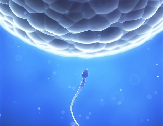
The first days after fertilization, when a sperm cell meets an egg, they are covered in a scientific mystery.
The process of how a humble single cell becomes an organism fascinates scientists in all disciplines. For some animals, the entire process of cell proliferation, create specialized cells and organize them into an arranged fetus multicellular takes place in the uterine protective environment, making immediate observation and provocative studies. This makes it difficult for scientists to understand what can go wrong during this process and how specific risk factors and the environment can prevent embryos from forming.
Scientists at UC Santa Cruz were able to build embryo cell models without ever experimenting with any real embryos, allowing them to imitate in the first few days after two sexual reproductive cells. They use CRISPR -based mechanical methods to push stem cells to organize “programmable” embryonic structures, also known as embryonic, which can be used to study the role of certain genes in early development. These structures are not truly embryos, but laboratory cells that are self-organized in ways that mimic certain aspects of early developmental stages. Their results are published in the leading magazine Stem Cell Journal Cellular cell.
We as scientists are interested in recreate and redefine natural phenomena, such as the formation of an embryo, on the plate to allow studies that otherwise provocative with natural systems. We want to know how cells are organized into a fetus -like model and what could go wrong when there are pathological conditions that prevent the success of an animal from successful. “
Ali Shariati, Assistant Professor of Biomological Engineering and Senior Author of the Study
Cellular co-development
Shariati is a specialist in stem cell engineering, a field that uses stem cells – non -specialized cells that can form any type of cells such as bowel or brain cells – to study and solve biological and health problems.
This project, led by UCSC’s postdoctoral researcher Gerrald Lodewijk and the biomolecular engineering graduate and current postgraduate student Caltech Sayaka Kozuki, used stem cells usually cultivated in the lab to guide them to form.
The team used a version of CRISPR technology known as an epigraine processor, which does not cut DNA but instead modifies how it is expressed. They targeted areas of the genome known to be involved in the development of an early fetus. This allowed them to control which genes were activated and to cause the creation of main types of cells required for early growth.
“We use stem cells, which are like a canvas empty, and use them to cause different types of cells using CRISPR tools,” Lodewijik said.
This method had the advantage of allowing different types of cells in “co-development”, which is more like the formation of a natural fetus than chemical approaches that other scientists have used to develop different cell types.
“These cells grow together, as in a real fetus and prove that the history of neighbors,” Shariati said. “We do not change their genome or expose them to specific signaling molecules, but rather activate existing genes.”
The group found that 80% of stem cells are organized into a structure that mimics the most basic form of an embryo after a few days and most are subjected to gene activation that reflects the development process that occurs in living organisms.
“The resemblance is remarkable in the way cells are organized, as well as the molecular composition,” Shariati said. “[The cells require] Very few inflows of us – it’s like cells already know what to do and we give them a little guidance. ”
The researchers noticed that the cells showed a collective behavior in movement and organization together.
“Some of them are starting to make this rotational immigration, almost like the collective behavior of birds or other species,” said Shariati. “Through this collective behavior and immigration they can form these fascinating fetal patterns.”
“Programmable” models
Having an accurate basic line model that reflects the early fetus of living organisms, it could allow scientists to study and learn how to treat development disorders or mutations.
“These models have a fuller representation of what is happening in the early stages of development and can capture the background,” Lodewijik said.
CRISPR programming not only allows scientists to activate genes at the beginning of the experimentation process, but also allows them to activate or modify genes significantly for other developmental parts. This allows embryo models to be “programmable”, which means that they can be relatively easily affected by a high level of control to target and control the effect of multiple genes as the fetal model develops, illuminating which have harmful results when activated or turned off.
For example, researchers have shown how some tissues are shaped or obstructed during early development, but their methods could be used to study a wide range of genes and their cataracts in cell types.
“I think this is the innovative work of this study – programming and that we are not based on exogenous factors to do so, but they probably have a lot of control over the cell,” Shariati said.
Researchers are interested in how this approach can be used to study other species, allowing a look at their embryo formation without ever using their real embryos.
This research could allow the study of congestion points that lead to reproduction to fail in the early stages. Among mammals, people have more challenges of reproduction in the fact that human embryos often fail to implant or determine the right early organizational form. Understanding why this is happening could contribute to progress to improve human fertility.
Source:
Magazine report:
Lodewijk, ga, et al. (2025). Self-organization of fetal mice fetal stem cells in reproductive embryo models before gastronomy through CRISPRA programming. Cellular cell. doi.org/10.1016/j.stem.2025.02.015.
