Discover how CKMT2 controls energy balance and mitochondrial health in skeletal muscle, revealing a new link between metabolism and type 2 diabetes management.
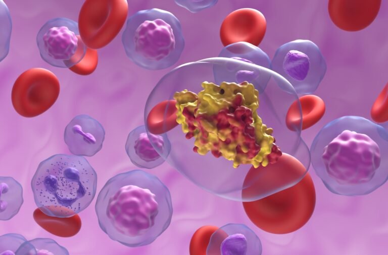
Study: Reduced mitochondrial creatine kinase 2 impairs skeletal muscle mitochondrial function independent of insulin in type 2 diabetes. Image credit: Nemes Laszlo / Shutterstock.com
A recent one Science Translational Medicine Study reveals that creatine kinase M2 (CKMT2) mediates mitochondrial dysfunction associated with type 2 diabetes.
The role of creatine kinases in type 2 diabetes
Altered energy metabolism occurs in patients with type 2 diabetes with insulin resistance due to limited intracellular storage and transport of adenosine triphosphate (ATP). Under these conditions, tissues that require high energy, such as skeletal muscle and the brain, use creatine kinases to convert creatine to phosphocreatine.
Creatine serves as an energy transporter due to its ability to store and transport ATP across the cell membrane. Creatine metabolism is associated with various pathophysiological functions, including immune responses in macrophages.
In fact, impaired phosphocreatine metabolism in white adipose tissue is associated with the development of a pro-inflammatory environment found in obese individuals. In men, elevated levels of circulating plasma creatine indicate an increased risk of type 2 diabetes.
Several studies have shown that changes in the expression and activity of creatine transporter proteins, as well as the interconversion of creatine to phosphocreatine, affect creatine metabolism.
Typically, creatine kinases are tissue specific with a unique intracellular localization. For example, the M-type isozyme of creatine kinase is localized in the cytoplasm of heart and skeletal muscle, whereas sarcomeric mitochondrial CKMT2 is located in the mitochondrial transmembrane space.
To rapidly hydrolyze ATP to adenosine diphosphate (ADP) for creatine phosphorylation, CKMT2 functionally co-localizes with the adenine nucleotide translocator (ANT). Although some studies have shown that CKMT2 is a vital regulator of mitochondrial respiration and oxidative phosphorylation (OXPHOS) in skeletal muscle, the functional role of CKMT2 in skeletal muscle metabolism in type 2 diabetes remains unclear.
About the study
The current study evaluates to identify any modification in CKMT2 content and creatine metabolism occurring in the skeletal muscle of men with type 2 diabetes. To this end, creatine metabolite levels were assessed, together with the expression of related genes from plasma samples and biopsy.
The role of CKMT2 was also assessed using mouse models fed a high-fat diet. In addition, the effects of Ckmt2 expression on mitochondrial metabolism were determined in C2C12 myotubes.
After clinical screening, 27 men with normal glucose tolerance and 25 men with type 2 diabetes were recruited. Participants with diabetes were treated with glucose-lowering drugs, including metformin and/or sulfonylurea. All drugs were taken after skeletal muscle biopsy samples were collected.
Study findings
Higher circulating fasting creatine levels were observed in plasma samples from men with type 2 diabetes and were negatively correlated with solute creatine transporter family 6-member 8 (SLC6A8) expression in skeletal muscle. Decreased levels of phosphocreatine were also observed, along with increased intramuscular creatine content, both of which were associated with CKMT2 expression in skeletal muscle.
Decreased skeletal muscle CKMT2 messenger ribonucleic acid (mRNA) levels were associated with higher post-oral glucose tolerance test (OGTT) circulating insulin in men with normal glucose tolerance and higher hemoglobin A1c (HbA1c) in men with type 2 diabetes.
CKMT2 expression was positively correlated with hip diameter in men with normal glucose tolerance. Except for glycine, which was decreased in men with type 2 diabetes, other creatine precursors remained unchanged in plasma. Body mass index (BMI) values did not influence creatine/phosphocreatine or gene expression in skeletal muscle.
Male mice were fed a high-fat diet for eight weeks, followed by treatment with phosphate-buffered saline (PBS), creatine, or the creatine analog β-guanidinopropionic acid (β-GPA) for the last two weeks of the diet. Although the high-fat diet increased mouse BMI, fasting glucose and glucose tolerance were not affected in these mice. Reduced basal and insulin-stimulated glucose transport was observed in the footpad of mice consuming a high-fat diet compared with those on a chow diet.
Mice treated with β-GPA showed improved basal glucose transport in the footpad. Creatine treatments partially reversed the high-fat diet-induced downregulation of CKMT2 mRNA. In addition, creatine treatment up-regulated genes encoding nitric oxide synthase 1 (NOS1), catalase (Cat), superoxide dismutase 1 (SOD1), nitric oxide synthase 3 (NOS3), hexokinase 2 (HK2), vascular endothelial growth factor factor (VEGF) and pyruvate dehydrogenase kinase 4 (PDK4).
Intact C2C12 myotubes were assessed via high-resolution respirometry analyzes on an Oxygraph-2k. To this end, CKMT2 silencing led to an overall reduction in mitochondrial respiratory capacity, as well as lower levels of hydrogen peroxide in myotubes and reduced mitochondrial membrane potential.
CKMT2 overexpression prevented lipid-induced metabolic stress in C2C12 myotubes as evidenced by oleate- and palmitate-induced downregulation of peroxisome proliferator-activated receptor γ coactivator 1α (Ppargc1a). This overexpression in skeletal muscle enhanced mitochondrial respiration and attenuated p38 mitogen-activated protein kinase (MAPK) activation in mice fed a high-fat diet.
Physical activity increases CKMT2 content, as twenty-five days of voluntary wheel running increased CKMT2 in a supercluster independent of mitochondrial biogenesis.
conclusions
The current study provides evidence for the role of CKMT2 in skeletal muscle mitochondrial homeostasis. Reduced CKMT2 expression may alleviate the main features of skeletal muscle dysfunction in patients with type 2 diabetes, including reduced mitochondrial function, reduced glucose metabolism, and increased reactive oxygen species (ROS) production.
Journal Reference:
- Rizo-Roca, D., Guimaraes, DSPSF, Pendergrast LA. et al. (2024) Reduced mitochondrial creatine kinase 2 impairs skeletal muscle mitochondrial function independently of insulin in type 2 diabetes. Science Translated Medicine. doi:10.1126/scitranslmed.ado3022.
