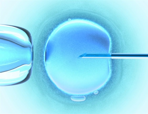
Researchers at the Catalonian Institute of Biomedicine (Ibec) in collaboration with Dexeus University Hospital have recorded unparalleled images of human embryo implant. This is the first time the process has been recorded in real time in 3D.
Failure to implant the uterus is one of the main causes of infertility, representing 60% of spontaneous abortions. Until now, this procedure could not be observed in real -time humans and the limited information available came from immovable images taken at specific times during the process.
“We noticed that human embryos are sinking into the womb, exerting significant power during the process. These forces are necessary because the embryos must be able to invade the uterine tissue, to be fully incorporated with it. It is an amazingly invasive process. Although it is known that many women experience abdominal pain and slight bleeding during implantation, the procedure itself had never been observed before “ Explains Samuel Ojosnegros, Ibec’s biomedical researcher for the reproductive health team and leader of the study.
To proceed during implantation, the fetus releases enzymes that break down the surrounding tissue. However, it is also known that power is needed to penetrate the underlying layers of the uterus. This fibrous tissue is full of collagen, a rigid protein that also forms tendons and cartilage. “The fetus opens a path through this structure and begins to form specialized tissues associated with the mother’s blood vessels to feed“He adds ojosnegros.
The results of the research team reveal that human embryos exercise attraction forces in their environment, reshaping it. ‘We observe that the fetus pulls the uterus of the uterus, moves and reorganize it. It also reacts to external signs of power. We assume that in vivo contractions may affect the implantation of the fetus“Explains Amélie Godeau, a researcher in the Ojosnegros team and the first author of the study. Thus, the effective invasion of the fetus is linked to the optimal uterine displacement, emphasizing the importance of these forces in the implantation process.
Improving the understanding of the implantation process could have a significant impact on fertility rates, fetal quality and time required to conceive through assisted reproduction.
A platform for the laboratory implantation study
To conduct the study, Ibec’s research team developed a platform that allows the embryos to implant outside the uterus under controlled conditions. This allows for real -time fluorescence to be depicted and analyzing the fetal interactions with its environment. The platform is based on a gel consisting of an artificial uterus formed by collagen, which is abundant in the uterine tissue and various proteins necessary for the development of the fetus.
Experiments were carried out with both human and mouse embryos to allow the comparison of the two implantation processes. When the mouse fetus comes into contact with the uterus, it exerts forces to adhere to its surface. The uterus then adapts to the folding around the fetus, the environment in a cache of the uterus. On the contrary, the human embryo moves inward and completely penetrates the tissues of the uterus. As soon as there, it begins to grow radially from the inside out.
‘Our platform allowed us to quantify the dynamics of embryo implantation and determine the mechanical footprint of the forces used in this complex process in real time“Concludes Anna Seriola, researcher of Ibec and co-authored author of the study.
This study was conducted in collaboration with Dexeus University Hospital (which donates all the embryos used in this study), biomimetic systems for the IBEC cell group, led by Elena Martínez, as well as other institutions such as The Insite Centers and Center Center (UB). in the biomedical (IRB Barcelona).
Source:
Magazine report:
Godeau, al, et al. (2025). The adhesion force and mechanical sensitivity are mediated by the patterns of implantation of special species in embryos and mice. Scientific progress. doi.org/10.1126/sciadv.adr5199.
